Name this rhythm!!

Handsome Robb

Youngin'
Premium Member
9,736
1,174
113
51 year old, trached, vent dependent male in an acute care SNF. Changing his shiley tube and he vomited and aspirated. Normally only on pressure support but they had to sedate him, suction the crap out of him and put him on SIMV to keep him saturating well.

Not really a scenario, more just looking for opinions on the rhythm. It was one of those calls that I didn't feel like I did well on. Nabbed the IV of a lifetime on the third attempt, 22g in the pointer finger between the first and second knuckles (proximal to distal) that flowed like a champion once I splinted that and the adjacent finger together with the help of a tongue depressor. Started 150 of amio over 10 minutes but were at the ER before it was finished, doc continued though so that made me feel a little more confident about my decisions.

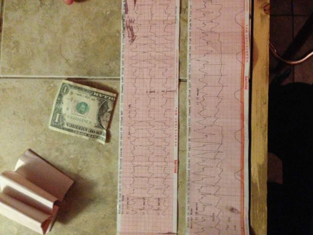
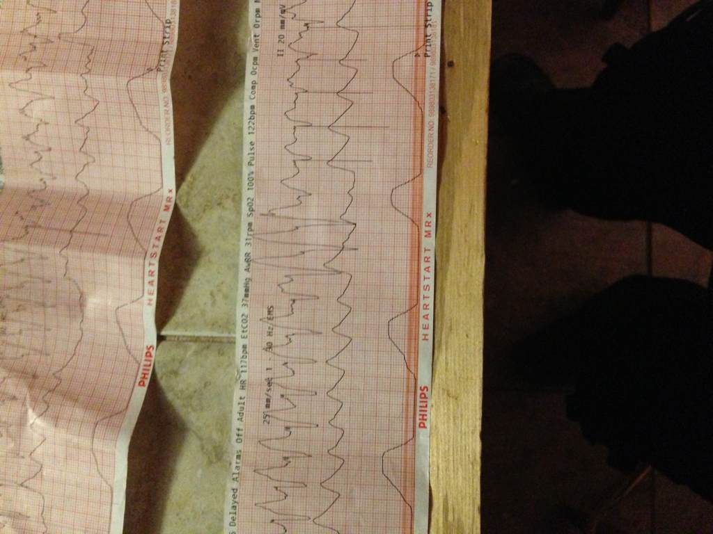
Sorry the really good pictures got turned in with my chart but this is the leftovers, should've printed everything again.
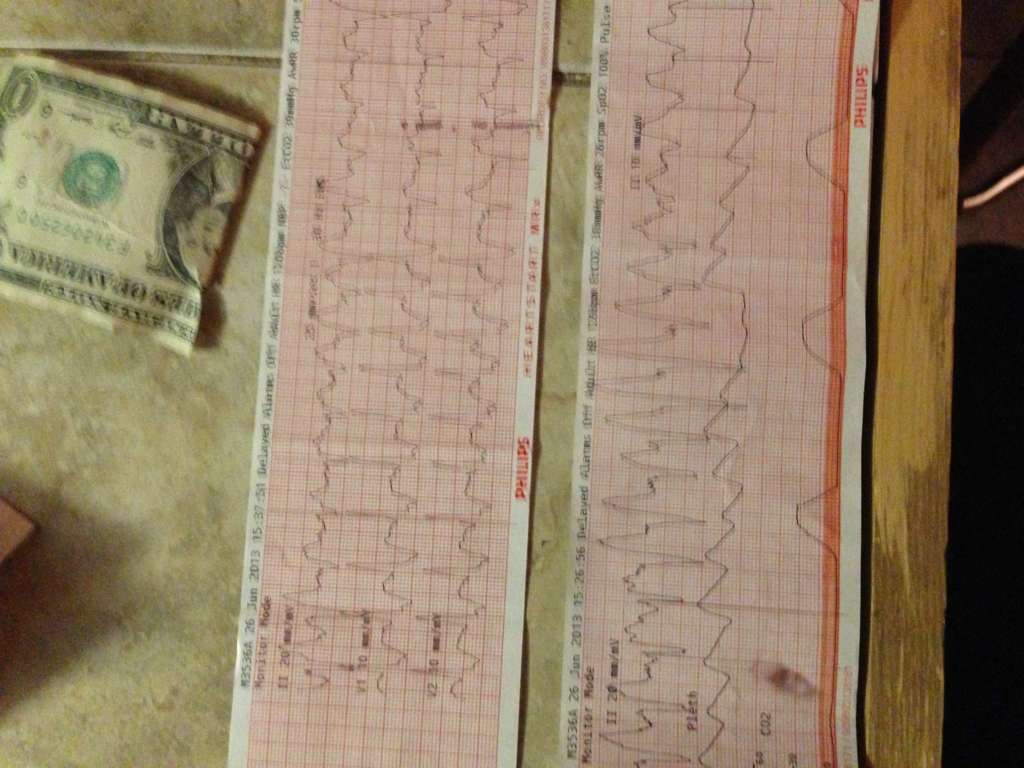
a6y5aqa6.jpg


u7u4yvyp.jpg


a2a9arej.jpg
 

VFlutter

Flight Nurse
3,728
1,264
113
Looks ugly. In the 12 lead I can see retrograde P waves buried in the T wave so Junctional with a conduction delay and Ventricualr arrhythmia are on my Ddx. Also Polymorphic ventricular ectopic beats with runs of VT.

Did he have a pacemaker? Looks like there is some pacing with various degrees of capture.
 

chaz90

Community Leader
Community Leader
2,735
1,272
113
I'd pretty much agree with Chase, minus the fact that I don't know where he found the 12 lead. No chance you have one of those floating around is there? I imagine you may have been somewhat preoccupied with other tasks.

BTW, I've found the CamScanner app works pretty well if you want to take pictures of paper EKGs.

Haha, I also almost asked if you guys got nasal prong EtCO2 or if you had the guy intubated after I saw the capnography waveform . Took me a couple moments to remember he was already trached and on a vent.
 
Last edited by a moderator:

VFlutter

Flight Nurse
3,728
1,264
113
I'd pretty much agree with Chase, minus the fact that I don't know where he found the 12 lead.

Oops no 12 lead. I meant the first strip with V1/V2
 

Aprz

The New Beach Medic
3,031
664
113
Looks like junctional tachycardia to me. There are associated p-waves after the complexes, and the complexes actually don't look that wide to me. Definitely a scary looking strip.

Interesting things about this strip in my opinion are:
- The R/S >1 in V1. Don't have I and V6 to look at for slurred S-waves to call it an RBBB. Doesn't look like RBBB to me in V1.
- The QRS axis changes in the precordial leads.
- When the axis changes, there is clearly no RBBB. Maybe rate dependent BBB?
- II is negative so the patient could have left axis deviation (more likely) or extreme right axis deviation (unlikely).
- Definitely has a ventricular pacemaker.

Since you have V1 and V2, I assume you did take a 12-lead, but you don't have it with you. :[
 
OP
OP
H

Handsome Robb

Youngin'
Premium Member
9,736
1,174
113
12-lead disappeared unfortunately. Ill see if I can pull the chart and download the ECG file that has the good strips and the 12-lead.

It was really regular but with the QRS volume cranked up you could hear the irregularity in it but there really wasn't a ton.

The picture with v1-2 was printed right before I did the 12 lead. Had to change monitor paper in the middle of the call too to add another aspect to an already difficult call.
 

Christopher

Forum Deputy Chief
1,344
74
48
12-lead disappeared unfortunately. Ill see if I can pull the chart and download the ECG file that has the good strips and the 12-lead.

It was really regular but with the QRS volume cranked up you could hear the irregularity in it but there really wasn't a ton.

The picture with v1-2 was printed right before I did the 12 lead. Had to change monitor paper in the middle of the call too to add another aspect to an already difficult call.

Paced ryhthm with lil runs of VT and some fusion with the pacer. Given the rate of the paced rhythm is 100 and that the VT doesn't inhibit it, seems like there is some sorta sensing failure/loop going on because the QRS-Pacer interval is far shorter than your typical fallback rate.
 
Top