Had a call for shortness of breath, ran the 12 lead and got this. Gimme what you got

Hockey

Quackers
1,222
6
38
I consider myself decent with 12 leads and I am always looking to learn something I may miss or don't know

This is one I had the other night.

Called for shortness of breath at like 430 in the morning at a nursing home.


85ish year old female. Recent CVA in July. Had shortness of breath about 40 minutes ago, nursing home gave a breathing treatment and took care of patients shortness of breath problem. No more wheezing. Patient has no complaints other than chronic back pain.

Hx: Hypertension, Diabetic II, CVA July with left sided deficits. No cardiac history besides hypertension. No lung problems other than "seasonal allergies".

The nursing home just got the patient about 24 hours prior. Nurse was useless as usual.

Lung sounds clear.

BP 152/98
HR 110-130
Resp 24
Pupils fine
Denies chest, head, neck and back pain.
Denies any shortness of breath
AO x 3 or 4


iXqg1.jpg


No old 12 leads available.

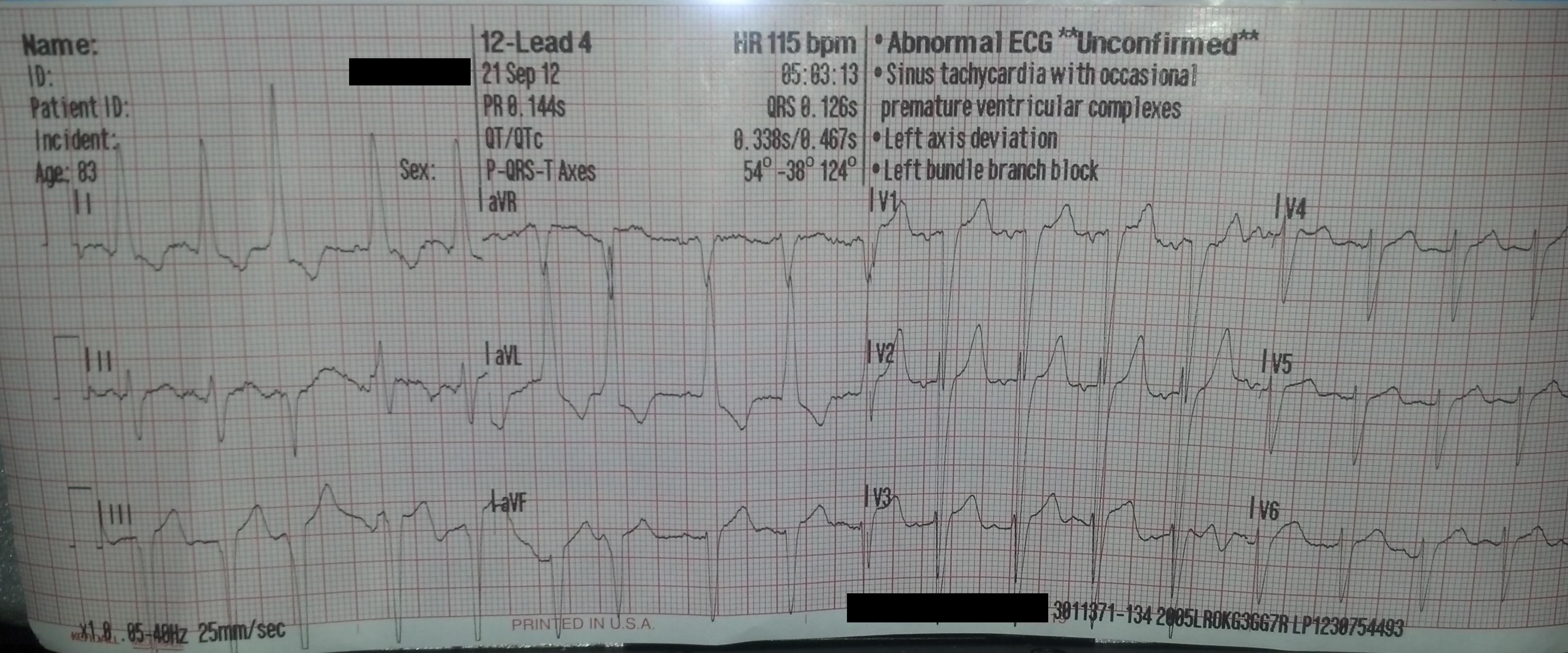
When we arrived at the hospital, the MD was waiting at the desk and I gave him a copy of the 12 lead. He decided since there was no old 12 lead available, and family was unable to be contacted, to activate a "Level II Acute MI"

Presence of left axis deviation. I see some elevation as well.

Just need to reassure some things about BBB's...when checking for BBB's, you look in V1 and if the QRS is greater than 120ms then it is, correct? I understand there should be a RsR wave in V6 but I don't see it here. Am I missing something?

What else do you see could be going on?
 
Last edited by a moderator:

NYMedic828

Forum Deputy Chief
2,094
3
36
She just had an exacerbation of some form of acute breathing episode, and was administered a beta agonist to resolve it. So, 115bpm especially with all the excitement doesn't seem too crazy.

May be nothing wrong and it is a chronic LBBB. QRS length is usually 120-140ms depending on the text you base the criteria on. Not sure about the v6 RSr'?


There is no presence of any concordant complexs. It also doesn't seem to meet Sgarbossa's criteria anywhere. (but someone told me that isn't very accurate?)

The best way to know, is to find out what the outcome was. (and let us know of course!)

I'm not that spectacular with 12 leads. I'm sure Chris or someone will eventually give you a cardiologist worthy answer.
 
Last edited by a moderator:

blindsideflank

Forum Lieutenant
184
3
18
Fast
Looks irregular
Left axis
P waves present, (except PVC) left atrial hypertrophy in V1
How many PVC in a minute? Are they really PVC or junctional with a conduction delay or atrial with the p hidden on the previous complex? Well it's different then qrs so ventricular.
Pr interval is good
Q nothing significant seen
Qrs wide, terminal S in V1 (QS or is it an rS?)
St segment- elevation seen is probably due to large qrs
Qt nothing significant

Sorry I'm on my truck riht now, I'll look at it better not on my iPhone.
Note, I am not stating facts, I'm thinking out loud
 

ChorusD

Forum Crew Member
40
0
6
Im saying it looks like left ventricular hypertrophy. LVH can apparently cause that STEMI mimic but I hear it can also cause that secondary depolarization abnormality that's manifested with a widened QRS angle.

Or...I could be way out in left field and she's having the big one. But I get most of those ideas from reading Tom Bouthillet's blog and others like it. Even though their blog posts are not text books I still think they are an outstanding resource for people like us.


And is it just my little iPad or did something happen to the picture? I could see the 12 lead before but it's gone now.
 

Sublime

LP, RN
264
6
18
Just need to reassure some things about BBB's...when checking for BBB's, you look in V1 and if the QRS is greater than 120ms then it is, correct? I understand there should be a RsR wave in V6 but I don't see it here. Am I missing something?

What else do you see could be going on?

You don't have to always have an RsR pattern in V6 to have a LBBB.

V1 is the main lead to look at, you're correct, but you need to look at the whole picture also. V6 is the second most important lead in BBB's, but its appearance can vary. Typically the terminal deflection of V6 should be opposite of V1, but again it can vary in some cases.

There should be no Q waves in the lateral leads in a LBBB.

You will most likely see some inverted, deep and slurred t waves in the lateral leads as well.

To me the 12-lead looks like a LBBB. According to her history and the lack of an old ECG it can be assumed this is a new onset LBBB which gets her a trip to the cath lab.

Here is some good videos on reading LBBBs.
http://ekgumem.tumblr.com/
Go to the bottom of the page. The last video is about diagnosing MI's in LBBB but he goes over briefly the criteria for a LBBB.

http://www.ecgteacher.com/index.php/conduction/bundle-branch-block
This video is more focused of recognizing the pattern associated with BBB's. It goes into detail and even has pictures and demonstration of the depolarization.
 

TomB

Forum Captain
393
82
28
Thanks, ChrorusD!

You are correct. Both LBBB and LVH with strain create a widened QRS/T angle (a general pattern of discordance) and this should always make you pause and think about it before declaring something to be a STEMI.

Negative concordance in the precordial leads (not referring to the pattern of T-waves opposite QRS this time but the fact that all QRS complexes are negatively deflected) is an unsual finding in LBBB. So I would check for a pacemaker pocket (always a good idea especially when you have a LBBB with left axis deviation although modern multi-site pacers create all kinds of patterns).

Assuming no pacemaker, this is why I use leads V1 and lead I to differentiate RBBB/LBBB/ICVD. Sometimes you can have a persistent S-wave in lead V6 and this is a normal variant. However, I do not allow this finding in lead I (which would make it a nonspecific intraventricular conduction defect). In other words, you should not have RBBB in the limb leads and LBBB in the precordial leads. When you do, you need to start thinking about the possibility of hyperkalemia.

Tom


Im saying it looks like left ventricular hypertrophy. LVH can apparently cause that STEMI mimic but I hear it can also cause that secondary depolarization abnormality that's manifested with a widened QRS angle.

Or...I could be way out in left field and she's having the big one. But I get most of those ideas from reading Tom Bouthillet's blog and others like it. Even though their blog posts are not text books I still think they are an outstanding resource for people like us.


And is it just my little iPad or did something happen to the picture? I could see the 12 lead before but it's gone now.
 
OP
OP
Hockey

Hockey

Quackers
1,222
6
38
I got first dibs on Toms brain when he dies.

Seriously.

I have learned more about 12 leads/EKG's from you and several others on the internet, and the Page class, than I ever did in any of my classes I have ever taken.
 

mycrofft

Still crazy but elsewhere
11,322
48
48
youngfrank64.jpeg
 

NYMedic828

Forum Deputy Chief
2,094
3
36
Thanks, ChrorusD!

You are correct. Both LBBB and LVH with strain create a widened QRS/T angle (a general pattern of discordance) and this should always make you pause and think about it before declaring something to be a STEMI.

Negative concordance in the precordial leads (not referring to the pattern of T-waves opposite QRS this time but the fact that all QRS complexes are negatively deflected) is an unsual finding in LBBB. So I would check for a pacemaker pocket (always a good idea especially when you have a LBBB with left axis deviation although modern multi-site pacers create all kinds of patterns).

Assuming no pacemaker, this is why I use leads V1 and lead I to differentiate RBBB/LBBB/ICVD. Sometimes you can have a persistent S-wave in lead V6 and this is a normal variant. However, I do not allow this finding in lead I (which would make it a nonspecific intraventricular conduction defect). In other words, you should not have RBBB in the limb leads and LBBB in the precordial leads. When you do, you need to start thinking about the possibility of hyperkalemia.

Tom

So what are we declaring this ECG to be?

I assumed it to be LBBB, unrelated to her condition.

You said it is unusual to have negative concordance in all precordials. In my experience v1-6 gets progressively more positive by v6 but I know it's possible to have all precordials be predominantly negative. I assume you are saying all negative concordance leads you to suspect pacemaker. But if you can't find physical evidence of a device (though the new ones are pretty dang small) can you presume it to be a LBBB if the rest fits?


Also, what you say about lead I having a prominent S wave opposite a normal lead I prominent R wave, what makes that suggestive of hyperkalemia? I know at a minimum it likely suggests right axis.
 

TomB

Forum Captain
393
82
28
When you have a wide QRS (= or > 120 ms) with LBBB morphology in lead V1 (QS or rS complex) you should have a monomorphic R-wave in lead I. In other words, you should have no S-wave at all. Not a little one. Not a prominent one. If you do it's not a LBBB, it's a nonspecific IVCD, the most life-threatening cause of which is hyperkalemia (if you don't consider it you can't diagnose it). It is VERY unusual for LBBB to show right axis deviation. If I saw LBBB with right axis deviation I'd make good and certain I was truly dealing with a supraventricular rhythm. I've seen one or two examples in my entire career (in textooks -- for example Chou has one example) but never in real life. LBBB should have a positive QRS in lead I and negative QRS in lead III.

It is also unusual for LBBB to have negative concordance across the precordial leads. In fact, with a WCT it's one of the ways we "rule-in" VT. A ventricular paced rhythm is a technical ventricular rhythm, which is why it's not unusual to show negative concordance across the precordials. Some might call this particular 12-lead a nonspecific IVCD because of the negative concordance in the precordials. However, the (somewhat arbitrary) criteria I use for LBBB uses leads V1 and I (again, because sometimes there can be a persistent S-wave in lead V6 although not always with a negative QRS).

Getting back to hyperkalemia, it is the "great imitator" but usually presents with nonspecific IVCD whent it gets to life-threatening levels. Often with QRS duration > 180 ms (but not always).

Tom

So what are we declaring this ECG to be?

I assumed it to be LBBB, unrelated to her condition.

You said it is unusual to have negative concordance in all precordials. In my experience v1-6 gets progressively more positive by v6 but I know it's possible to have all precordials be predominantly negative. I assume you are saying all negative concordance leads you to suspect pacemaker. But if you can't find physical evidence of a device (though the new ones are pretty dang small) can you presume it to be a LBBB if the rest fits?


Also, what you say about lead I having a prominent S wave opposite a normal lead I prominent R wave, what makes that suggestive of hyperkalemia? I know at a minimum it likely suggests right axis.
 
Top