Interesting EKG

chaz90

Community Leader
Community Leader
2,735
1,272
113
I'm happy to give a scenario and/or more background information later, but I initially want to see what everyone thinks of these recently acquired EKGs.

mezuqusa.jpg


2u9abady.jpg


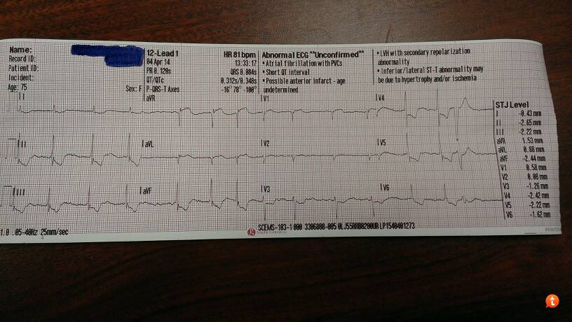
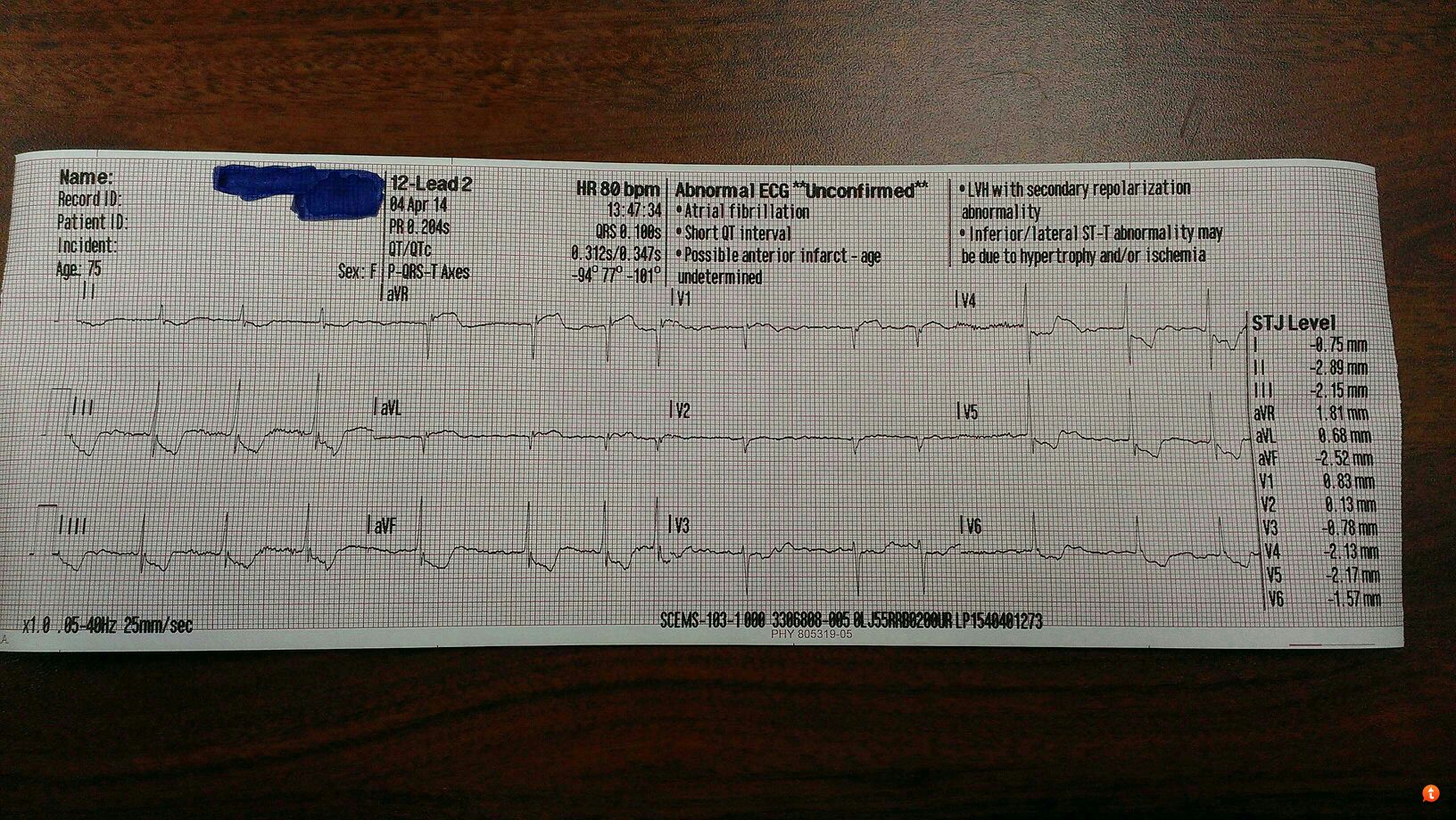
Anyone have any thoughts?
 

Handsome Robb

Youngin'
Premium Member
9,736
1,174
113
Someone needs a cath lab now. Like right now. Probably a CABG in all reality. just looking at the ECG without knowing anything else I'd say proximal LMCA occlusion...

STE in aVR + STE in aVL + ischemic changes.
 
Last edited by a moderator:

TomB

Forum Captain
393
82
28
I can't disagree. It must be correlated to the history and clinical presentation but I've seen very similar ECGs with ROSC patients who ended up with 3-5 vessel bypasses (after cath showed severe multi-vessel disease).
 
OP
OP
chaz90

chaz90

Community Leader
Community Leader
2,735
1,272
113
It must be correlated to the history and clinical presentation

And there unfortunately is the rub in this case. This was my partner's call, so I only saw initial contact with the patient prior to them transporting and then discussed the EKG and treatment with her after she returned from transporting. 75 YOF from a SNF, AMS progressing over the last several days after discharge from hospital for "encephalopathy." We asked for more details regarding the patient's baseline mental status and what type of encephalopathy but were unable to obtain any from nursing home staff.

Pt was unresponsive for us the entire contact. No stated complaints according to staff, and they related it was more of a gradual change over the past three days rather than anything acute. Staff denies history of dementia and believes she is there for rehab only with a goal of discharge to home and independent living. Staff relates history of UTI and significant cardiac problems but are unable to provide additional details.

Initial BP 132/72, HR 80 (a-fib with history of same), SpO2 of 94% with a good waveform, RR 20, EtCO2 35 and non-obstructed. Axillary temp of 99 F (potentially unreliable LP15 probe) and BGL of 360 mg/dL. GCS of 6 (E:1 V:2 M:3). LS clear, pupils equal, and no other significant physical exam findings.

12 lead EKGs as shown above, and my partner wished to begin treating the patient with NTG paste only as she was clearly unable to take SL NTG or ASA. Before paste able to be applied, pt. grew hypotensive (80s systolic) and was treated with ~750 mL NaCl with an ending pressure at destination of 100/60. No change in responsiveness or other significant changes in VS. No POC lactate reading available unfortunately.

I called the ED later for follow up and was told the patient was admitted with a diagnosis of urosepsis and encephalopathy (again, unknown further details). The nurse said their 12 lead showed similar changes to ours but that her troponin was negative and they weren't working her up for any suspected cardiac issues.

I still thought this was an interesting EKG and I at least grew more familiar with some of these characteristic findings of STE in aVR. Thanks for playing team!
 

teedubbyaw

Forum Deputy Chief
1,036
461
83
What's the significance of the aVR elevation? Any other teaching points on this one?

I'm pretty good at 12 lead interpretation at my level, but this one is another league.
 

VFlutter

Flight Nurse
3,728
1,264
113
Likely Toxic/Metabolic Encephalopathy with urosepsis. Unless there is a history of hepatic or renal failure. My guess is the EKG represents Demand Ischemia in the setting of sepsis, and likely anemia, with significant multivessel CAD. History of Diabetes? If not, stress hyperglycemia. I would think this is the early stages of what will end up as SIRS/Sepsis.
 
Last edited by a moderator:

Summit

Critical Crazy
2,694
1,314
113
I'm not the strongest at 12 lead but the avr/avl st segments simply match a global ischemic picture.

Unresponsive with those vitals doesn't match an AMI

You don't have an indication for nitro here I don't think!

You have unexplained mental status changes, somewhere between marked and extreme since you don't know baseline but you know its not what you are looking at for a patient aiming at rehab and d/c to independence. You have an older lady with global ischemia, unresponsive, high glucose, and a history of UTI.

What is always high on the differential for old ladies with a primary presentation of confusion/AMS? UTI.

What do we not typically see until late if at all in geriatric sepsis? Fever

... but you would expect ischemia (demand).

The bolus was a good choice. The ntg could have been a disaster! You got this patient right at the transition from sepsis to septic shock.
 
Last edited by a moderator:

medicsb

Forum Asst. Chief
818
86
28
Initial BP 132/72, HR 80 (a-fib with history of same), SpO2 of 94% with a good waveform, RR 20, EtCO2 35 and non-obstructed. Axillary temp of 99 F (potentially unreliable LP15 probe) and BGL of 360 mg/dL. GCS of 6 (E:1 V:2 M:3). LS clear, pupils equal, and no other significant physical exam findings.

Was she really posturing?
 
OP
OP
chaz90

chaz90

Community Leader
Community Leader
2,735
1,272
113
I suppose we could upgrade the GCS to 7 with a motor score of 4. Seems more appropriate for her.
 

Brandon O

Puzzled by facies
1,718
337
83
It's not a CLASSIC ECG for intracranial insult (honking inverted T waves), but just about any ECG changes are technically consistent with noggin troubles. You could even argue for something like Tako-Tsubo. However, as stated, demand ischemia coupled with widespread baseline stenosis is probably the most plausible.

Echo would make things a little clearer in the acute setting (as would obtaining her records on cardiac hx), but the negative troponin works.
 

Handsome Robb

Youngin'
Premium Member
9,736
1,174
113
Hmm. Learn something new every day! With that story I definitely agree that ACS this is not.
 

Christopher

Forum Deputy Chief
1,344
74
48
Without remembering any specific criteria I thought possible bleed. Ill look into specifics in a sec here (assuming the case was odd and it wasn't left main).

http://lifeinthefastlane.com/ecg-library/raised-intracranial-pressure/

Example #2 from the above link

The original ECG is distinctly different from neurogenic T-waves seen during high catecholamine load.

The QTc is much shorter in chaz's ECG, along with the classic ACS style ST morphology found in an anatomical distribution.

Anywho, chaz's ECG is much more likely due to shock.
 

MSDeltaFlt

RRT/NRP
1,422
35
48
No STEMI. ST segment elevation and/or depression also includes the S wave of the same complex. Which is not happening in this case.
 

Psych

Forum Probie
18
0
0
You have a pretty typical case for advanced urosepsis here.

I had a patient very similar to this, call came out of a SNF, she was A&Ox0-1 (baseline A&Ox4), having convulsions, being treated for urosepsis with levoquin (due to her PCN allergy...terrible decision, they should have been using aztreonam - levoquin alters coumadin levels and is associated with an increased risk of seizures in the elderly). The differences was my patient has elevated troponin and had a pacemaker. Elderly women are susceptible to urosepsis already, Hx of DM2 makes complications much more likely. Your patient was exhibiting clear multi organ dysfunction associated with the sepsis - she probably was deteriorating in septic shock as well. The encephalopathy is not a coincidence, it was sepsis associated (http://www.ncbi.nlm.nih.gov/pubmed/22986430). I wouldn't be surprised to learn of some kind of sepsis associated DIC or a significant thromboembolic event was on the horizon for this patient.

Anytime there's an increased metabolic demand on the body some arrythmia is probably going to show up. My patient was in AFib with paced ventricular rhythm, developed up to 6 second runs of PVCs before transport, deteriorated into VTach/VFib (perhaps pacemaker induced?) and did not make it. She was also in respiratory distress, and I think there were vegetations on the pacer leads showering emboli into her lungs (the leads are normally on the RV septal wall). Really just poor performance on the part of her doctor, who allowed her to be discharged from the ED and come back to the SNF after the Dx so he could treat it (poorly, with the wrong abx) and probably result in her death. Wow I let this turn into kind of a rant, well hopefully it helps someone understand this case which seems to be pretty common.

EDIT: Is it just me or can you only see PVCs in the first 12-lead in V4-6?
 
Last edited by a moderator:

TheLocalMedic

Grumpy Badger
747
44
28
Alright, hold the phone... This seems like a case of ignorance, if you want my opinion.

Please tell me that you weren't thinking AMI because aVR and aVL were showing some elevation. I just had a similar ECG presented to me where a medic was concerned that their "STEMI" patient wasn't sent to the cath lab despite elevation in aVR and aVL. The confusion lay in the fact that they thought aVR and aVL were "continuous leads", whereafter I had to explain to them that, despite the leads being displayed next to each other they were not, in fact, contiguous leads, and that there was little correlation between the two.

I had to have a whole sit-down teaching session where I explained the schematic...

I Lateral aVR V1 Septal V4 Anterior
II Inferior aVL Lateral V2 Septal V5 Lateral
III Inferior aVF Inferior V3 Anterior V6 Lateral

Although the leads appear next to each other, they do not necessarily look at the same parts of the heart.
 

VFlutter

Flight Nurse
3,728
1,264
113
I had a patient very similar to this, call came out of a SNF, she was A&Ox0-1 (baseline A&Ox4), having convulsions, being treated for urosepsis with levoquin (due to her PCN allergy...terrible decision, they should have been using aztreonam - levoquin alters coumadin levels and is associated with an increased risk of seizures in the elderly).

Since when is aztreonam available PO? Unless she had an IV at the SNF. I rarely see it used to begin with. Levequin is an appropriate treatment for a UTI. The risk of seizures is minimal and really only a major concern in patients with preexisting seizure histories. Levaquin can result in supratheraputic INR when used in patient's on coumadin but is usually not an issue in the relatively short time they are on an antibiotic regime.

Your patient was exhibiting clear multi organ dysfunction associated with the sepsis - she probably was deteriorating in septic shock as well. The encephalopathy is not a coincidence, it was sepsis associated (http://www.ncbi.nlm.nih.gov/pubmed/22986430). I wouldn't be surprised to learn of some kind of sepsis associated DIC or a significant thromboembolic event was on the horizon for this patient.

Where do you see clear mutliorgan dysfunction? I see AMS. I wouldn't consider hyperglycemia. Is increased metabolic demand and ischemia an organ dysfunction? That is pushing it. Depending on white count, which I assume is elevated, she barley meets SIRS criteria. She is non-tachy, non-hypoxic, and not hypotensive. I do not think she was in deteriorating septic shock. Will it end up there? Most likely. But I have seen numerous patients with significant AMS from infections without sepsis and shock.
 
OP
OP
chaz90

chaz90

Community Leader
Community Leader
2,735
1,272
113
I didn't believe the patient was suffering from an AMI. I absolutely agree that the presentation was completely consistent with sepsis, and I buy demand ischemia as the likely source of the EKG changes. Again, my partner transported this patient, and no STEMI alert was called, nor was any NTG used.

I'm not ignorant enough to believe aVR and aVL are contiguous, and it wasn't just that that made me do additional research on this EKG. As mentioned earlier in the thread by some people with fairly extensive EKG background, these kind of changes can frequently be found in patients with LMCA occlusion, proximal LAD occlusion, and triple vessel disease. This case didn't represent a cardiac issue, but STE in aVR>STE in V1 and aVL along with diffuse ST depression have been associated with the above changes.
 

jrm818

Forum Captain
428
18
18
Since someone asked earlier about avr as well, here's some background reading.

Obviously the rest of the scenario is wrong for ACS, as I think everyone recognized , but given that ECG with the right story, "uh oh...me think you need cabg....me drive to hospital with cath lab and surgeon" might be the right thought, even though avr and avl are indeed not anatomically contiguous.

http://ekgumem.tumblr.com/post/17948970835/lead-avr-deserves-your-respect-episode

http://content.onlinejacc.org/article.aspx?articleid=1127536

http://hqmeded-ecg.blogspot.com/2011/04/st-elevation-in-avr-with-widespread-st.html

http://lifeinthefastlane.com/ecg-library/lmca/
 

Brandon O

Puzzled by facies
1,718
337
83
The whole aVR thing is tricky. We should all be grateful to (primarily) Dr. Mattu for popularizing its association with widespread or LMCA occlusion, but some of the data is not totally convincing about the predictive value.

My solution has been in the clinical correlation -- if they look sick as :censored::censored::censored::censored:, widespread depression (rarely elevation... total LMCA occlusions are usually codes), elevation in aVR, give it some thought. But hopefully none of you knuckleheads were going to call that pericarditis anyway.
 
Top