EKG Interpretation

chaz90

Community Leader
Community Leader
2,735
1,272
113
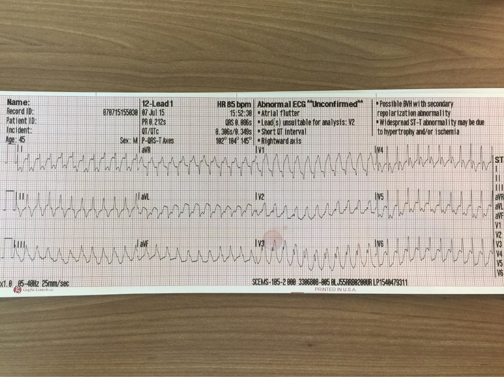
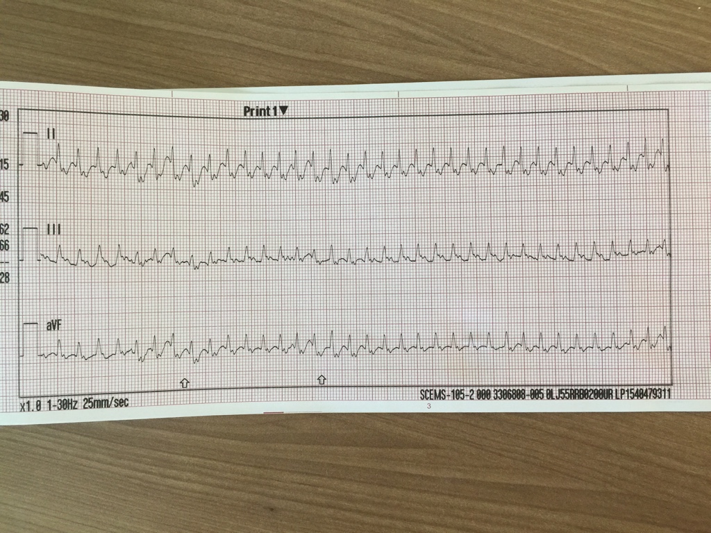
Here's one for the EKG buffs. This is an absolutely cold read, as my partner and I literally found this EKG in the trash can at our station this AM. We know it's a 45 year old male as that information was input to the monitor, but we have no clinical scenario, symptoms, history, or vitals of any kind. My partner and I interpreted this rhythm differently, so I'd love to see a little discussion about it on here. Without further ado, here's the single 12 lead we have and a rhythm strip.

6b157f02cdc6ff68c700a5cf6315daaf.jpg

8f42a2d57c359460908353791f74be90.jpg
 
OP
OP
chaz90

chaz90

Community Leader
Community Leader
2,735
1,272
113
I now found out a little bit more about the treatment and outcome, so I'll post those results after I hear some thoughts.
 

Brandon O

Puzzled by facies
1,718
337
83
Was it actually a 45-year-old male or was that just a default?
 
OP
OP
chaz90

chaz90

Community Leader
Community Leader
2,735
1,272
113
I believe it ended up actually being a 53 year old male
 

Aprz

The New Beach Medic
3,031
664
113
I chuckled a little when I saw that monitor said the rate was 85. The rate is clearly close to 300.

It is a regularly regular narrow complex tachycadia. I believe it is an SVT.

Types of SVT
- sinus tachycardia
- atrial fibrillation
- Atrial flutter
- AVNRT
- accessory pathway mediated tachycardia
- atrial tachycardia
- multifocal atrial tachycardia
- junctional tachycardia

I kinda forget the order of most to least common, but it is something like that.

The whole 220-age for upper sinus tachycardia rate is a good guideline, but not a rule. This is obviously well beyond even 220... probably not sinus tachycardia.

The rhythm looks regularly regular to me, but it is possible that the rate is making it look regular. At such a high rate it is hard to tell sometimes. I would say about 300 is still too high for atrial fibrillation.

Oddly, I think the monitor may be right about atrial flutter even though it is totally wrong about the rate. The rate is just about right for an atrial flutter rate 300 (I consider around 250-350 normal. Some literature say 200-400). Since this is about 300 that would make 1:1 atrial flutter pretty high on my differential. This would also explain the very odd looking ST changes and concordancr of those changes because the flutter waves would be superimposed on top of the complexes and T wave. The oddball thing to me is where I see the flutter waves best. Typically atrial flutter is best seen in the inferior leads (II, III, and aVF) and lead V1. I also usually see it in lead aVR (due to it being opposite of lead II). On this ECG, I see it on almost every lead.

Note atrial flutter and atrial tachycardia are pretty similar, but the atrial rate is slower (150-250 instead of 250-350). Both can have variable blocks which is not present here.

AVNRT is still a possibility. The most common form (slow-fast) will have an pseudo s wave in III and pseudo RBBB or pseudo r wave in lead V1. The less common types of AVNRT will make a double hump in the T wave or even look like sinus tachycardia with a 1st degree block.

At a rate of 300, an accessory pathway mediated tachycardia is definitely a possibility.

Like atrial fibrillation, multifocal atrial tachycardia is an irregular rhythm.

Junctional tachycardia is the rarest making it least likely. This goes up my differential if the patient is taking digoxin.

Another possibility is VT (it looks wide in some leads and the widest lead, longesr QT, longest PRi, etc. are what you use). I believe the leads that are wide are due to the ST changes or superimposed flutter waves. A narrow form of SVT is fascicular VT. It'll have a RBBB morphology, which this does not have, and the most common form of fascicular VT has left axis deviation, which this does not have. This is more inferior axis or around +90 degrees.

There is really nothing else I can add in regard to interpretation. I would call it an SVT. I think 1:1 atrial flutter is most likely.

I would follow AHA guidelines. If the patient was hemodynamically stable then I would establish and line and administer fluids. I would attempt to the have the patient vagal down if the IV didn't do the trick. I would administer adenosine. If that didn't work then I would sedation with synchronized cardioversion. I would be concerned about the monitor approproately recognizing the QRS complexes.

If he was hemodynamically unstable then I would immediately do synchronized cardioversion. 1:1 atrial flutter is not tolerable for long and the patient will probably be hemodynamically unstable. Arrial flutter does respond well to electricity I believe.

If we didn't go all the way to cardioversion and the patient was stable, then I would expect the hospital to administer a Ca2+ channel blocker like diltiazem or cardiovert the patient themselves.
 

TomB

Forum Captain
393
82
28
The relatively young age and extreme rate suggest WPW Syndrome. Could be a reciprocating tachycardia or 1:1 flutter. You could apply the defibrillator pads and attempt adenosine which would either break the rhythm or confirm 1:1 flutter. Or, if hemodynamically unstable, sedate and perform synchronized cardioversion.
 

Aprz

The New Beach Medic
3,031
664
113
I beat TomB by 1 minute to pressing the post button. Woo...
 

Christopher

Forum Deputy Chief
1,344
74
48
1:1 atrial flutter tops my list, although AVRT is a possibility. The neat bit about 1:1 flutter is it doesn't actually need an accessory pathway to conduct that quickly as certain conditions can "enhance" AV nodal conduction (e.g. high sympathetic tone, hyperthyroid).

I've got a nearly identical 12-Lead in my collection, with only minor differences actually!
 
OP
OP
chaz90

chaz90

Community Leader
Community Leader
2,735
1,272
113
Good info from all! Not surprisingly, all three of you mention the correct diagnosis. This ended up being 1:1 atrial flutter. The treating medic recognized it as a narrow complex regular tachycardia, though did not initially suspect AF. He treated the patient (after placing defib pads) with the standard 6 mg followed by 12 mg doses of adenosine which broke the rhythm down to atrial flutter 2:1 with incredibly obvious flutter waves and an immediate change to a rate of ~144. Patient exhibited stable vitals signs with a CC of chest pressure and a "racing heart" and the transporting medic ran out of time to administer Diltiazem before they arrived at the hospital.

I'll admit, I initially suspected VT when looking at this EKG. Looking back on it now, the flutter waves are fairly apparent as Aprz noted and I should have caught those. I think I was fooled into thinking the QRS was wider than it truly was by looking at some of the buried p-waves. I recognized the axis deviation as being unusual for VT but didn't want to rule it out on that alone.
 

Brandon O

Puzzled by facies
1,718
337
83
The rate is the most helpful aspect here IMO. Once you're this fast you have to argue me out of flutter.

But really the point is that it doesn't matter. Treatment will be the same for all of these.
 

Handsome Robb

Youngin'
Premium Member
9,736
1,174
113
Damnit I was too slow. With the rate my first thought was A-flutter.
 
Top