ECG Scenario. VT or SVT with Aberrancy?

Handsome Robb

Youngin'
Premium Member
9,736
1,174
113
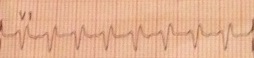
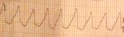
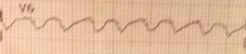
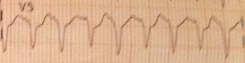
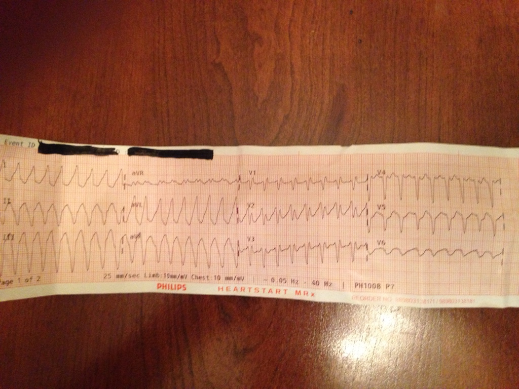
58 year old male presents with non-radiating/reproducible retrosternal chest pressure ranging from a 4/10 to 8/10 with associated SOB and diaphoresis. Started 30 minutes after a 'normal' hour long workout. Reduced with rest then increased 15 minutes after taking the trash down a flight of stairs to the dumpster and returning to his apartment which happened 5 initial onset. PT has had similar episodes over the last 3 months with 'normal' echocardiogram and stress test within the last 4 weeks.

PT is GCS 15 cool, pale and diaphoretic. Physical exam is otherwise unremarkable.

Vitals:
112/90
HR as seen at 210 with corresponding pulses.
96% on room air
RR 34 with clear lung sounds bilaterally.

History:
HTN
MI "a few years ago" with stent x2 in RCA
Chronic back pain

NKDA

Meds:
Lisinopril
Flexeril
Hydrocodone/APAP

Non-smoker, rare social ETOH, no drug use

12c2ced8065aa3114e615cc9d64450d6.jpg
 

DesertMedic66

Forum Troll
11,273
3,452
113
I would call this SVT with aberrancy. However I always have a hard time telling the two apart.
 

TomB

Forum Captain
393
82
28
Robb:

The ECG shows clear-cut VT.

The desire to call a wide complex tachycardia SVT with aberrancy is very powerful. I'm not entirely sure why but it's true! One of my missions in life is to counteract this desire in the minds of emergency clinicians.

Here are some things to keep in mind:

  • The history MI makes VT very likely in this case.
  • VT is far more common than SVT with aberrancy.
  • "Wide and fast" is the most important criterion for VT.
  • The burden of proof is on the person who claims a rhythm is SVT with aberrancy (and the evidence should be quite compelling).
  • The morphology here is not typical for RBBB or LBBB.
  • Various algorithms like Wellens and Brugada have demonstrated poor inter-rater reliability (although in this case they strongly favor VT).
  • Failure to rule-in VT does not rule out VT (it need not have a right superior axis, positive or negative concordance in the precordial leads).

Fortunately, the distinction of VT vs. SVT with aberrancy is not necessary to treat the patient appropriately! It's enough to know it's a wide complex tachycardia but not so wide that it's suspicious for hyperkalemia.

Tom B.
 

Clare

Forum Asst. Chief
790
83
28
This is VT. We are taught a wide complex tachycardia you cannot obviously differentiate from VT must be treated as VT until proven otherwise.

Hmm, expeditious transport. If close to hospital never mind calling for ICP and just take him. If a bit further away I would seek clinical advice regarding the appropriateness of cardioversion. At Paramedic level cardioversion is possible but it is "awake" cardioversion so not the nicest for pt but an alive pt with a sore chest is still better than a pt in cardiac arrest.
 

chaz90

Community Leader
Community Leader
2,735
1,272
113
Hmm...I'd interpret this as VT although without all the reasoning Tom B was able to provide!

Going off that, close monitoring of BP and mentation, 150 mg amiodarone drip, and place the multi-function pads on the patient's chest. I'd draw up 0.1 mg/kg Etomidate with plans to administer it IV if cardioversion becomes necessary.

Treatment for me would depend on transport time too. I would absolutely start the amiodarone regardless of distance to the hospital, but if the amiodarone drip failed to convert his rhythm and he remained symptomatic over the course of an hour long transport, I'd consult with med control about proceeding with cardioversion despite his currently stable BP and mental status. SOB, chest pressure, and diaphoresis with a HR of 210 in a 58 year old with history of MI is pretty dang concerning to me, and I wouldn't love keeping this rhythm going for any longer than I had to.
 

Aprz

The New Beach Medic
3,031
664
113
We already talked about this, but I'll repeat what I said.

The rate is totally prime for ventricular tachycardia (VT). I think most literature probably says around 150-250 beats per minute. I estimated the rate to be around 187-214. You mentioned that the heart rate was 210.

It's obviously a regularly regular wide complex tachycardia. This is where we come up with our differential of VT (most common) or supraventricular tachycardia (SVT) with aberrancy (usually due to a bundle branch block or an pathway mediated tachycardia such as antigrade atrioventricular re-entrant tachycardia (AVRT)). Without even looking at the ECG, but being told it is a regularly regular wide complex tachycardia, you will likely be right if you blindly guess VT. It's gonna be VT the majority of the time.

A lot of interpreters use Brugada's criteria to look for specific findings that would favor VT vs SVT with aberrancy.
  • AV disassociation
  • Lack of RS complexes in precordial leads
  • Beginning of R-wave to nadir of S-wave >100 ms
  • Atypical bundle branch block morphology.
Those are all findings that favor VT.

As soon as I saw this ECG, I felt that the morphology was atypical for bundle branch block. Intermediate interpreters are gonna look at lead V1 to figure out if it is closest to right or left bundle branch block. They are gonna look at I and V6 to support that it is a bundle branch block. If I and V6 are not correct then they might call it an intraventricular conduction delay (IVCD) which is most commonly caused by hyperkalemia.

v7BLuEB.jpg

V1​

This looks like an rSR' OR Qr, which closest mimics RBBB. RBBB usually has a qR or rsR'. (I am so used to typing rsR' and qR that I totally messed that up when I talked to you privately... I saw your confused reply when you said "but RBBB is rsR' or qR", haha.) I don't consider this very typical looking for RBBB though even though it is close notation wise. It just doesn't look right.

RBBB will usually have a Rs "slurred S" wave in I and V6.

BdqSzYm.jpg

I​

Lead I in your ECG is totally not normal for RBBB under any circumstance that I can think of. There is clearly no slurred S wave in it.

NElTXfm.jpg

V6​

I talked to you about RBBB with left axis devation (LAD), left anterior fascicular block (LAFB), or together called a bifascicular block. I've noticed that I and V6 (usually V6 more in my opinion) will have more of an RS wave (deeper S-wave pretty much) in a bifascicular block. Although this looks like RBBB with left axis deviation, V6 doesn't really look like what I am describing. RBBB with LAD in a wide complex tachycardia is pretty suggestive of VT. Not very likely to be a tachycardic bifascicular block.

The other issue is that the complexes are very wide, especially in the frontal axis. The wider it is, the more likely it is VT.

That's why I believe this is considered to be atypical bundle branch block morphology.

I am personally not a fan of looking for AV disassociation unless it is really obvious to me. Although specific, it is not super sensitive. I have a hard time distinguishing artifact vs AV disassociation. It should like changing morphology (especially the T-waves where "hidden" p-waves from AV disassociation) throughout the lead(s) it is present in. If I am not mistaken... I think an argument could be made that AV disassociation is present in lead V5, but I always think it is possible because of poor image quality or artifacts, which is common in the prehospital setting.

pzYmeqw.jpg

V5​

My #1 rule with differentiating between VT vs SVT with aberrancy is that the ECG must be very clear cut. I don't try to go all trick like "it could be a SVT with a bifascicular block." It has to be perfect. Even then... some VTs can present with perfect RBBB or LBBB morphology because of where the foci is. Some VT can have narrow (even narrower than SVT with aberrancy) complexes (eg fascicular block). We talked about the possibility of this being a fascicular block (commonly RBBB with LAD), but this is too wide in my opinion to be a fascicular VT. We talked about right ventricular outflow tract (RVOT) VT, which would show as a RBBB with inferior axis (or towards the II, III, and aVF making those three leads totally positive and the mean QRS vector axis to be around +90). This is definitely not RVOT VT since the axis it is left axis deviation (I is positive. II, III, and aVF are negative. aVR is the smallest making the mean QRS vector perpendicular to that making it around -60 degrees).

As others have mentioned, the patient's age and history (previous MI) support ventricular tachycardia.

I totally believe this is ventricular tachycardia. I would consider this patient to be hemodynamically unstable because of the chest pressure, shortness of breath, and poor skin signs. I would have considered sedation (versed and morphine) since his blood pressure was good, it appears his symptoms have been lasting several hours, and he was alert and oriented I assuming. It would be a nice thing to do, but I really would be leading towards immediate cardioversion still. I am not a fan of chemical cardioversion.
 

Brandon O

Puzzled by facies
1,718
337
83
It's always VT. It's always VT. It's always VT.

I would consider cardioversion, because I want to see a more readable QRS/T complex. I'm worried about that RCA.
 

Aprz

The New Beach Medic
3,031
664
113
Life in the Fast Lane also mentions the morphology of I (and V6).

http://lifeinthefastlane.com/ecg-library/basics/right-bundle-branch-block/

They have at the bottom of their web page
We invite you to use our content in anyway to help others learn, all we ask is that you spread the word about the FOAM (Free Open Access Meducation) revolution...and get #FOAMed !
Click the links for information on FOAM if you aren't familiar with it.

The following image is from Life in the Fast Lane.

Broad-S-wave-RBBB-ann-590x209.jpg

Normal Lead I for RBBB​

Lead I and V6 will look very similar.

http://lifeinthefastlane.com/ecg-library/basics/bifascicular-block/

In their example on bifascicular block, you'll see that V6 doesn't have that typical Rs wave and doesn't look like lead I. It's more of an RS wave.

I am just trying to explain why your ECG doesn't demonstrate typical RBBB morphology.
 

bizzy522

Forum Crew Member
61
9
8
I look at this and think VT. ACLS wide tachycardia algorithm says for hymodynamicly stable patients to consider adenosine first then follow amiodarone. I always thought it was to rule out a possible aberrancy if one was unsure?
 

zzyzx

Forum Captain
428
90
28
Don't over think it. He's getting cardioverted right now. Considering how he looks and what he's complaing off, it's a simple decision.
 
Top